Retinal Tear
A torn retina is a serious sight-threatening problem that can cause symptoms like seeing flashes of light, new black dots, moving cobwebs or “floaters” across your vision. Without prompt treatment, a torn retina often leads to a retinal detachment, where the retina is lifted away from the back of the eye resulting in severe vision loss. Urgent eye review and treatment of retinal tear is required and when retinal laser treatment is provided promptly in the clinic, retinal detachment and surgery can be prevented.
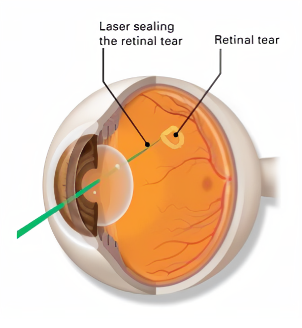
Image from American Academy of Ophthalmology: Cataract Surgery
How does a torn retina occur?
The retina lines the back wall inside the eye and it is a delicate layer of nerve cells critical for our eye sight. A tear or hole in the retina can occur due to a number of factors including ageing changes of the eye, as a result of eye injury or surgery, or in highly short-sighted eyes. As we get older, the vitreous jelly that fills the middle of our eyes and is immediately adjacent to and supports the retina, starts to shrink and lose its protective firmness. As the vitreous jelly degenerates and shrinks, in healthy ageing, it normally separates away from the retina without causing any problems except for some floaters. However, in some instances, the vitreous jelly maybe more “sticky” to the retina and instead of separating away cleanly, it can pull hard enough away from it to tear it. When that happens, fluid (liquified vitreous) can pass through the tear and lift or detach the retina, leading to vision loss. A detached retina can no longer function normally, resulting in vision loss and blindness.
Who is at risk of developing a torn retina?
People are at most risk of developing a torn retina around the ages of 40-50 years when their vitreous jelly undergoes age-related degenerative changes as just described above. However, the age at when this occurs is highly variable and retinal tear can occur at younger and older age groups. Some of the individual risk factors that can put you at higher chance of developing a torn retina include:
- Being short-sighted (or needing highly myopic prescription glasses to see far away).
- Having a retinal tear or detachment in the other eye.
- Having family members who have had retinal detachment.
- Having weak or fragile areas in the retina (which your optometrist or eye specialist may find during a dilated retinal examination).
- Having had previous cataract, glaucoma or other eye surgery.
- Having had significant eye injury or trauma.
What are the symptoms of a torn retina?
A torn retina has to be urgently detected and treated by an ophthalmologist right away. Otherwise your retina could detach and you could lose vision in that eye. Urgently contact your eye specialist if you have any of the following symptoms:
- You become acutely aware of many new floaters (shaped like moving small dots, hairs, flies or cobwebs).
- You see flashing lights. These are often very brief flashes of light (like a camera flash) and are most noticeable in the dark or dim illumination.
- A shadow or gray curtain appears in your peripheral (side) vision.
How is a retinal tear diagnosed?
Only a thorough and detailed dilated retinal examination by an experienced retinal specialist can detect small or very peripheral retinal tears or holes. Your ophthalmologist will put drops in your eye to dilate (widen) the pupil. He will then look into the back of your eye through special optical lenses and use a diagnostic microscope to examine all the areas of your retina in 3D. Standard 2D imaging or retinal scans can easily miss a peripheral retina tear or hole. Also, a 2D wide field digital retinal imaging can also result in a false positive result, where a retinal tear or hole is incorrectly diagnosed when in fact, there is no such problem. Either way, it is vital to have the definitive diagnosis confirmed by your ophthalmologist, giving you the reassurance and confidence in the most appropriate treatment and/or monitoring plan being instituted.
How are retinal tears treated?
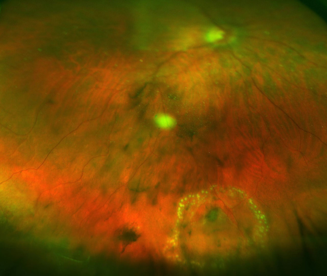
Clinical photo: Optos wide-field fundus imaging showing successful laser treatment of a peripheral retinal tear (at 6 o’clock position). The yellow dots correspond to the laser treatment sealing the tear (upside down “V-shaped” break).
Retinal laser is used to seal the torn retina and this can be done safely and comfortably in the eye clinic. The goal of the laser treatment is to permanently seal the retinal break and thereby prevent it enlarging or from fluid going through the tear and detaching the retina. Retinal tears or holes are usually treated on an urgent basis. This can protect your eye sight and also prevent the need for eye surgery. However, if a torn retina has unfortunately progressed to a detached retina, retinal surgery can be performed to re-attach the detached retina.
If you would like more information on the treatment options for retinal tears, please call Eastwood Eye Specialists for an appointment.
Monday - Friday
8:30 am to 4:30 pm
Saturday (once a month)
8:30am to Noon
© 2021- Eastwood Eye Specialists
Website by: ![]()